9) Empty the sac with a syringe and remove the cannula.
Arm Venipuncture, Injection, Transfusion (Blood Transfusion)
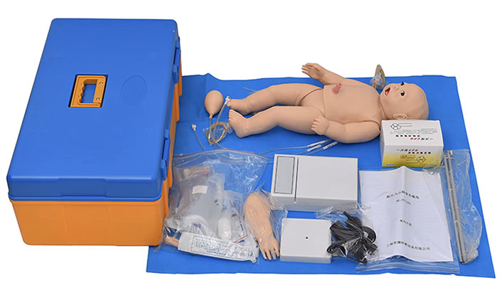
The arm venipuncture training model consists of a plastic upper limb arm showing the elbow forearm veins (cephalic vein and vital vein) and infusion vessels and other supporting devices, one of the most common teaching models for basic clinical nursing practice.
Training program
I. Blood sampling through elbow forearm vein
Intravenous injection or intravenous infusion through the elbow and forearm.
Trans-elbow forearm venous blood transfusion
1) Installation method:
Two rubber tube clamps were set into the plastic upper limb model connected to the two rubber tubes, a rubber tube up and through the connecting sleeve with the blood simulation fluid infusion bag (bottle) connected (Note: the beginning of the practice, the infusion bag first use water, proficiency and then use the blood simulation fluid. Preparation of blood simulating solution: it is recommended to use 4 grams of simulated blood powder and 100 ml of water to make the solution). The other rubber tube is down and inserted into the waste fluid bottle.
I. Blood is drawn through the forearm vein at the elbow:
Steps (1) 200-300 ml of blood simulation fluid connected to the plastic upper limb model upper rubber tube, so that the blood simulation fluid injected into and filled with the plastic upper limb model within the piping system, clamp the lower rubber tube clamp to block the lower rubber pipeline.
Step (2) Routinely sterilize the skin of the elbow forearm.
Step (3) Select a suitable vein, puncture the vein with a 10 ml syringe and withdraw 2 ml of venous blood (blood simulation fluid).
II. Intravenous injection or intravenous infusion through the forearm of the elbow
Intravenous injection or intravenous infusion is the pressurized injection of medicinal solution into a vein, commonly used veins are cephalic vein and noble vein.
Uses: (1) Rescue or treatment of patients by injecting glucose solution and medication or blood patients through pressurized veins, increasing the amount of blood in coronary arteries and carotid arteries so as to improve the blood in the heart and brain, and rescuing or treating the patients by reflexes to bring the blood pressure back up. (2) Used to perform certain special examinations. (3) For chemotherapy.
Intravenous injection or intravenous infusion training:
Step (1) is the same as step 1 of drawing blood through the forearm vein at the elbow, using a syringe tray, a suitable syringe, a No. 6-8 needle, medication, a sandbag, sterile gloves and sterile therapeutic wipes. This method of injection is contraindicated in patients with hematologic disorders, as it may cause hemorrhage.
Step (2) Routinely disinfect the skin of the elbow forearm with sterile gloves and sterile cavity towel.
Step (3) Fix the selected vein with the index and middle fingers of the left hand, hold a syringe (50 ml syringe, No. 6-8 needle) with the other hand to extract the medicinal fluid, stab the vein vertically or vein at an angle of 40 degrees in the direction of stabbing the vein, and then pull back to see the red liquid entering the syringe, clamp the upper rubber tubing clip to block the upper rubber tubing, loosen the lower rubber tubing clip, and then fix the puncture needle with one hand while pushing the medicinal fluid as fast as possible with the other hand. Push the medicine as fast as possible, so that the medicine in the syringe flows through the piping system in the model, so that the medicine enters the waste liquid bottle through the lower rubber tube. Remove the needle quickly when the injection is complete. Intravenous infusion when stabbed into the vein back to see the red liquid into the syringe, then release the rubber tube clamp, adjust the intravenous infusion device drip speed, so that the infusion bottle of red blood simulation liquid flow through the model of the piping system and through the lower rubber tube into the waste liquid bottle, fixed puncture needle.
Third, blood transfusion through elbow forearm vein:
Intravenous blood transfusion operation training
Step ① with the elbow forearm venous blood drawing step 1.
Step ② Routine sterilization of the skin of the elbow forearm.
Step ③ Select a suitable vein, puncture the vein with a hypodermic needle, pump back to see the red liquid into the venous transfusion device, adjust the drip speed, so that the red blood simulation liquid in the infusion bottle flows through the piping system in the model and through the lower position of the rubber tube into the bottle of waste liquid.
IV. Bone marrow aspiration and intraosseous infusion
Bone marrow aspiration training
Step 1: Place the model on top of the table, apply lubricating powder to lubricate the whole bone marrow puncture parts, install and slide the right tibia bone marrow puncture parts from the sole of the foot upward to the fixed position of the lower leg tibia, and cover the lower leg with a skin jacket.
Step 2: Place a disposable waterproof dust cloth pad that absorbs liquid under the knee joint of the model, connect the adjustable infusion frame, infusion bag and its connecting pipeline with the tibial bone marrow puncture component, install and adjust it to become ready for infusion, and eliminate air bubbles in the pipeline. (At the beginning of the operation, the infusion bag should be filled with pure water first, and then blood simulation liquid should be used after you are skillful in the operation. (Preparation of blood simulating solution: it is recommended to use simulated blood powder 4 grams plus 100 ml of water to prepare).
Step 3, (1) Check that the tibial bone marrow puncture component is filled with fluid. Prevent leakage of blood simulating fluid during operation.
(2) Bone marrow puncture, should use a special bone marrow puncture needle, from the skin to the periosteum for adequate anesthesia, forearm and bone marrow puncture needle as an axis, and make it turn back as the main points, for slow pressure into the bone marrow, to prevent excessive pressure, puncture correctly can be simulation of bone marrow liquid outflow. The sample can be taken with a 5 ml syringe. The bone marrow fluid extracted is generally 0.1-0.2 ml, if used for bacterial culture, 1-2 ml of bone marrow fluid can be extracted. (3) The tibia bone marrow puncture component is designed to puncture on all four sides, and after the bone marrow puncture operation, the bone hole is sealed with a small piece of wax. After one side of the puncture component has been punctured, the component can be rotated 90 degrees and reinserted into the tibia until all four sides of the tibial bone marrow puncture component have been punctured, at which time the component can be discarded.
Intraosseous infusion.
Intraosseous infusion: is the introduction of fluids, blood or medications directly into the tibial bone marrow or into other bones. Intraosseous infusion is an ancient technique that is fast, simple, safe, and effective in the clinical application of infant shock resuscitation, and is especially suitable for patients in whom vascular access cannot be established during resuscitation (when severe dehydration and blood loss are present, and peripheral veins are almost invisible or untouchable, and resuscitation access cannot be established).
Intraosseous infusion is mostly used in the tibial position. Method of bone marrow puncture in the right tibia of the simulator: asepsis is required to establish an inlet for intraosseous infusion. A bone marrow aspiration needle (16-gauge) is inserted 1 cm below the tibial tuberosity, pressurized and rotated back and forth to pierce the bone marrow aspiration needle to a depth of 2-3 cm, penetrating the bone cortex, there is a sensation of falling air, and the core of the needle can be withdrawn with a syringe to draw out the simulated bone marrow fluid. The intraosseous inlet is connected to the intraosseous infusion channel with a needle connector, and the needle connector is clamped with hemostatic forceps and secured to the leg; after stabilization, the intraosseous inlet can be used to inject fluids, blood, or medications. It is recommended in the literature that intraosseous infusions usually need to be performed for 1-2 hours until a safer venous access system has been established. Ensure that the fluid in the tube is drained after each use. Use glue or sealant to close the pinhole after an intraosseous fluid puncture to prevent fluid leakage. The tibial module can also be inverted for puncture or replaced with another new tibial module.
V. Femoral Artery Pulse Beat Examination Procedure:
Simulate the operation of femoral artery pulse measurement
1、Assist the patient to take the supine or sitting position, the arm is placed in a comfortable position and the wrist is stretched.
2、will be installed in the simulator of the human body on the left side of the plastic catheter and rubber pressure ball connection, hand pumping to simulate the femoral artery pulse (note the pulse rate, rhythm, strength).
3、Press the tip of the index finger, middle finger and ring finger on the surface of the femoral artery, the pressure is appropriate to be able to clearly touch the pulse.
4、Counting: Normal pulse is measured for 30S, multiply the number of measured pulses by 2 to get the pulse rate.
VI. Cardiopulmonary Resuscitation (CPR) First Aid Training
The Cardiopulmonary Resuscitation (CPR) monitor provides the correct cadence and monitors for proper blowing and compressions. There are catheter openings on the left side of the simulator's torso: a lung exhaust catheter opening, which connects to the blow detection port on the rear of the monitor; a cardiac exhaust catheter opening, which connects to the compression detection port on the rear of the monitor; and an arterial pacing balloon pumping catheter opening.
Implementation standards: The product adopts the American Heart Association (AHA) 2020 International Cardiopulmonary Resuscitation (CPR) and Emergency Cardiovascular Care (ECC) guideline standards.
Functional features:
1、Standard airway opening: tilt the head and lift the chin method
2、Support mouth-to-mouth, mouth-to-nose, simple respirator-to-mouth and other ventilation methods, electronic monitoring of blowing frequency, blowing volume, compression frequency and compression depth.
CPR is operated according to the ratio of compression and ventilation: compression: blowing is 30:2.
3、the whole operation timing function, the operation process can be a key to reset to start the next round of operation, the whole voice prompts, volume can be adjusted at will, in the process of operation can be a key to mute, the whole digital counting, compression and blowing the correct and incorrect number of times.
4、can be pressed on the simulator:
Pressing depth: about 4cm
Pressing frequency: 100-120 times / min
Depth indicator of compression status (right side of the human body picture on the monitor):
When the display is yellow, the pressing depth is insufficient. (Pressing error count 1 time)
When the display is green, the pressing depth is correct. (Pressing correctly counts 1 time)
When the display is red, the compression depth is too deep. (Pressure error count 1 time)
Blow volume indicator (left side of the human body picture on the monitor):
When the display is yellow, the blowing volume is insufficient. (blowing error count 1 time)
When the display is green, the blowing volume is correct. (correct blow count 1 time)
When the display is red, the blowing volume is too large. (blowing error count 1 time)
Cardiopulmonary resuscitation (CPR) demonstration procedure:
Installation: Separately connect the blowing (Vent) detection plastic catheter, pressing (Comp) detection plastic catheter and CPR external power adapter on the left side of the human body of the simulator with the interface on the rear side of the monitor, and then turn on the power button of the machine. Press the Start button to operate when the selection is complete.
Insufficient rebound voice alarm.
Artificial respiration (blowing) display alarm:
The green barcode light shows when the blowing volume is correct; the yellow barcode light shows when the blowing volume is too small and the voice alarm is insufficient; the red barcode light shows when the blowing volume is too large and the voice alarm is too large.
Pressing and manual blowing 30:2, complete 5 cycles of CPR operation.
1、Power supply: power switch
2、Operation Timing: operation time counting
3、Start:Start operation
4、Power supply: detect the power supply status
5、On-line:Detect the connection status
6、Volume:Volume up and down selection
7、Mute:Mute key
8、Frequency:frequency selection key (green light 100/min, red light 120/min)
9、reset: reset to the initial state
Simulator right interface description:
1) right arm infusion (shared) outlet (scalp vein and arm head vein)
2)Left arm infusion (shared) inlet (scalp vein and arm head vein)
3) Umbilical vein infusion inlet
4) Oral-nasal gavage gastric tube infusion outlet
5) Umbilical vein infusion outlet
6) Right foot infusion (shared) outlet (right marrow cavity infusion, right femoral vein and left saphenous vein)
7) Left foot infusion (shared) inlet (right bone marrow cavity infusion, right femoral vein and left saphenous vein)
Description of the interface on the left side of the simulator:
1) blowing
2) compression
3) Pulse beat pressure bulb connected to catheter inlet

Name: manager zheng
Mobile:0086-13588958091
Tel:0086-0577-62626932
Whatsapp:8613588958091
Email:sales@kangmugroup.com
Add:qiaoqian industrial zone, liushi wenzhou, zhejiang,325600,china
We chat
